A common complication of any wound healing process is the breakdown of tissues within and around the injury site. In milder cases, tissue death is minimal with little effect on wound healing but in more severe cases, tissue necrosis can lead to permanent limb loss and disability. In this article, we will describe some strategies for managing wound necrosis.
Defining Necrosis
Cell necrosis refers to the pathological death of a cell or group of cells following exposure to a prolonged, and harmful stimulus. Necrosis can be contrasted to the more orderly process of apoptosis, where cell death is triggered by regulatory signals sent to a specified group of cells.
What Is a Necrotic Wound?
Necrotic wounds are areas of tissue loss that occur following the death of their component cells. Once an area of tissue becomes devitalized, dead tissue build-up commences which might inhibit the rate at which wound repair occurs. Usually, the onset of necrosis can always be traced to the development of pathology or exposure to a harmful physical or chemical insult.
Pathophysiology: How Do Necrotic Wounds Form?
As mentioned earlier necrotic wounds result from various harmful stimuli which halt or alter the normal metabolic processes within human cells. In many cases, nutrient and oxygen deprivations due to a compromise of blood supply to the affected areas trigger death on a cellular level which later manifests as visible tissue necrosis.
Classifying Necrotic Wounds
Necrotic tissue can be categorized either as eschar or slough. While eschar is a thick, firm, and dark-pigmented necrotic material covering a wound site, slough is a softer, moist, yellow-to-tan material found within and around a wound’s margins.
Regardless of the type of necrotic material present at a wound site, the effect on tissue repair is the same. When allowed to remain at the wound site, necrotic tissues will form a physical barrier preventing optimal wound healing and providing an ideal environment for bacterial colonization and infection.
Etiology/Risk Factors
Various external factors predispose individuals to the development of wound necrosis. The most commonly cited causes of wound necrosis are:
- Trauma
- Infections (bacterial, fungal, or viral)
- Toxins (from animal and insect bites or stings)
- Tissue inflammation
- Cancers
- Vascular compromise (ischemia)
- Chronic disease
Diagnosis
Diagnosing necrotic wounds is a pretty straightforward process involving the identification of devitalized tissues (slough or eschar) around or within a wound site. Wound care professionals will typically combine physical wound examination with relevant patient history to diagnose, and treat necrotic wounds.
Treatment Strategies for Necrotic Wounds
Due to the negative effect necrotic tissues have on any wound healing process, including limb loss, and permanent disability, wound care experts must be proactive in their approach to treating these types of tissue injuries. The mainstay of necrotic wound treatment is local wound care with patients benefiting from other adjunct therapies including analgesia, antibiotics, exercise, and dietary modification.
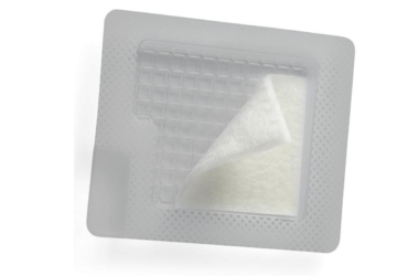
Local Wound Care Strategies
The most critical aspect of treating a necrotic wound involves dedicated wound care practices. Wound cleaning, debridement, and dressing will allow wound care providers to clear the site off devitalized tissues, preserve the function of affected limbs, and boost overall healing.
Types of Wound Debridement
Wound debridement can be achieved using the techniques listed below:
- Mechanical debridement
- Autolytic debridement
- Sharp debridement
- Surgical debridement
- Biological debridement
Mechanical Debridement
This involves applying moist-to-dry dressings onto the wound site. When peeled off, the necrotic tissue attached to the dressings is removed. The main drawback of this technique is the tendency to remove both dead cells as well as healthy granulation tissues.
Autolytic Debridement
Autolytic debridement is a moisture-enabled process that uses the fluid within the wound to break down and remove de-vitalized tissue. In some cases, the moisture required for autolytic tissue breakdown can be supplied from externally applied wound dressings.
Sharp Debridement
Sterile scissors or forceps can be used by wound care professionals to separate necrotic tissue from the wound site to allow for unhindered tissue repair. This technique requires the services of a wound care professional to prevent injury to vital structures within and around the wound site.
Surgical Debridement
This technique is done by a surgical wound care professional under sterile conditions with local or general anesthesia in an operating room. This technique is reserved for patients who have large areas of necrotic tissues that require more extensive debridement. In these cases, a surgical environment is mandatory to reduce the risk of bleeding and infection after the debridement process is completed.
Biological Debridement
Biological agents (larva or maggots) cultured in a sterile environment can be used to remove necrotic tissue from a wound site. The larvae secrete enzymes that break down the dead tissues which they then consume. The benefit of using biological agents is that the larvae do not attack healthy tissues which makes them very efficient in clearing the wound site. However, most patients find the idea of applying maggots to their skin repulsive so biological debridement therapy is usually a last resort.

Tags

.jpg)

.jpg)

.jpg)