Chronic wounds are a significant burden to the healthcare systems around the globe. In the United States, approximately $28 billion is spent annually on patients with chronic wounds. Chronic wounds are characterized by a prolonged inflammatory process which delays the overall wound healing process. Normal epidermal migration is impaired in chronic wounds which results in the accumulation of non-viable devitalized tissue in the wound bed. The presence of devitalized tissue in the wound bed impedes healing and acts as a nidus for wound infection. Therefore, early identification and management of devitalized tissue are important for the promotion of wound healing. Podiatrists and wound care specialists should be aware of the different types of non-viable tissue that might be present in chronic wound beds.
Types Of Devitalized Tissue
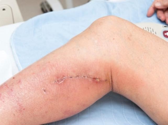
For optimum wound healing, the wound bed should be well-perfused and free from devitalized tissue. Therefore, wound bed preparation is an integral part of chronic wound care. To avoid the removal of healthy granulation tissue, it is important for podiatrists and wound care specialists to be able to correctly identify non-viable tissue in the wound bed. Broadly speaking, non-viable devitalized tissue can be classified into two main types:
- Eschar: It is dry, leathery necrotic tissue present in non-healing wounds. It is usually present in full-thickness wounds.
- Slough: It refers to different forms of nonviable devitalized tissue present in the wound bed. It usually appears as yellowish white material and is composed of leucocytes, macrophages, and fibroblasts. In addition, bacterial colonization results in a ‘biofilm’. It is essential to distinguish slough from healthy granulation tissue.
A new classification system has been proposed to define different types of devitalized tissue that might be present in wound beds:
- Necroslough: This type of devitalized tissue is black or brownish in appearance, and is usually situated over thin bony prominences. It results from a total lack of blood supply, and as result, becomes shriveled, hardened, and mummified. Incomplete removal of necroslough can trigger an inflammatory process at its base resulting in liquefactive necrosis.
- Leukoslough: Leukoslough results from infiltration of the wound bed with white blood cells. It appears as a soft, yellowish-white gelatinous material. It is loosely adherent to the wound bed and can be easily removed.
- Fibroslough: The deposition of fibroblastic collagen results in the formation of fibroslough. Even though it might be similar in appearance to leukoslough, it is more firmly attached to the wound bed.
- Bioslough: This type of slough results from the progressive accumulation of biofilm on the wound surface. It is dark maroon, greenish black with a foul odor. Antimicrobial dressings should be used to prevent the accumulation of biofilm.
Management Of Devitalized Tissue
The presence of devitalized tissue delays wound healing and should be removed for the progression of normal wound healing. Devitalized tissue can mask underlying abscesses and can make it difficult to determine the extent of the wound bed. Removal of necrotic tissue decreases bacterial load and infection rates which improve healing. The following management options are currently available for the management of devitalized tissue:
- Wound Irrigation: This involves the removal of surface contaminants, debris, and slough from the wound bed using normal saline. Low-pressure irrigation is suitable for the removal of loosely adherent material while high-pressure irrigation may be beneficial in highly-contaminated wounds.
- Surgical Debridement: It involves the surgical removal of dead, necrotic tissue using a sharp instrument. It is indicated for the removal of large areas of dead, necrotic tissue. Surgical debridement has been found to decrease the bacterial burden and significantly improves wound healing through stimulation of wound epithelialization. Surgical debridement requires a skilled practitioner, and might also be painful.
- Autolytic Debridement: It is a conservative method of debridement primarily used in patients with infected wounds. It occurs naturally in the wound bed by proteolytic enzymes and phagocytic cells. A moist wound environment facilitates autolytic debridement. Therefore, moisture-retaining wound dressings are indicated for this method of debridement. If no significant decrease is observed in the amount of devitalized tissue after a few days, an alternate method should be considered.
- Enzymatic Debridement: This involves the application of exogenous enzymes to the wound bed. Enzymatic debridement is a suitable alternative for patients who are not able to tolerate surgical debridement. Even though healing rates are not improved with the use of most enzymatic agents, collagenase may stimulate angiogenesis and endothelial cell proliferation which helps to accelerate wound healing.
Biological Debridement: Also known as “maggot therapy”, biological debridement involves the use of larvae for the debridement of devitalized tissue. Through the release of proteolytic enzymes, the larvae can digest and remove the necrotic tissue present in the wound bed. The use of maggot therapy has also been associated with a reduction in the use of antibiotics. It is a painless method and is suitable when large areas of devitalized tissue need to be removed

.webp)
.avif)