
Understanding the wound healing process is essential for healthcare professionals to develop appropriate treatment plans tailored to individual patients' needs. Different wounds may require different interventions based on their stage of healing and underlying causes. For instance, wounds with necrotic tissue, such as foot ulcers with necrosis, require debridement to remove dead tissue and promote healing. Similarly, wounds with signs of infection may require antibiotic therapy. By comprehensively understanding wound healing, healthcare providers can optimize patient care and outcomes.
What Is a Necrotic Wound?
Definition and Characteristics of Necrotic Wounds
Necrotic wounds refer to areas of tissue death or necrosis within a wound site. Necrosis can occur due to various factors, including ischemia, infection, trauma, or underlying medical conditions. In necrotic wounds, tissue becomes non-viable and appears black, brown, or dark. Necrotic tissue can impede wound healing by serving as a barrier to new tissue formation and promoting bacterial growth, leading to delayed healing and increased risk of complications.
Identification of Necrotic Tissue in Wounds
Identifying necrotic tissue within a wound is crucial for developing an appropriate treatment plan. Common signs of necrosis include discoloration of the wound bed, presence of eschar (dry, black, leathery tissue), and foul odor. Necrotic tissue may also feel firm or hard to the touch. Healthcare professionals assess wounds for necrosis during wound examination and debridement procedures. Prompt identification and removal of necrotic tissue are essential for facilitating wound healing and preventing further tissue damage or infection.
How Do Necrotic Wounds Form?
Mechanisms Leading to Necrosis in Wounds
Necrotic wounds can form through various mechanisms, often disrupting the normal wound-healing process. Ischemia, or inadequate blood supply to tissues, is a common cause of wound necrosis. This can occur due to vascular diseases, such as peripheral arterial disease, which restrict blood flow to the affected area. Infections can also contribute to necrosis by causing tissue damage and inflammation. Additionally, traumatic injuries, such as burns or crush injuries, can lead to necrosis by disrupting blood vessels and compromising tissue viability.
Factors Contributing to the Formation of Necrotic Tissue
Several factors can contribute to the formation of necrotic tissue within wounds. Prolonged pressure or friction on vulnerable areas, such as pressure ulcers or diabetic foot ulcers, can lead to tissue ischemia and necrosis. Inadequate wound care, including improper cleaning or dressing changes, can also predispose wounds to necrosis by promoting infection or hindering tissue repair. Underlying medical conditions, such as diabetes or peripheral vascular disease, can further increase the risk of necrosis by impairing circulation and compromising tissue health. Prompt identification of contributing factors and implementation of appropriate wound care interventions are essential for preventing or managing necrotic wounds effectively.
Causes of Necrosis
Underlying Conditions and Factors Leading to Necrotic Wounds
Peripheral Arterial Disease (PAD): PAD is a condition characterized by narrowing or blockage of the arteries that supply blood to the extremities, leading to decreased blood flow. Reduced blood flow deprives tissues of oxygen and nutrients, resulting in ischemia and tissue necrosis. In severe cases, PAD can lead to gangrene, particularly in the lower extremities.
Diabetes Mellitus: Diabetes can cause peripheral neuropathy and vascular complications, both of which increase the risk of necrotic wounds. Peripheral neuropathy diminishes sensation in the extremities, making individuals less aware of injuries or pressure points that could lead to tissue damage. Vascular complications, such as peripheral arterial disease and microvascular dysfunction, impair blood flow to tissues, exacerbating ischemia and tissue necrosis.
Traumatic Injuries: Trauma, such as burns, crush injuries, or severe lacerations, can cause direct damage to tissues, disrupting blood vessels and impairing blood supply. Inadequate blood flow to the affected area leads to tissue ischemia and necrosis. Traumatic injuries may also introduce pathogens, increasing the risk of infection and further tissue damage.
Infections: Bacterial, fungal, or viral infections can cause tissue necrosis by inducing inflammation, releasing toxins, and damaging surrounding tissues. Infections can compromise blood flow to the affected area and impair the body's immune response, allowing pathogens to increase and spread, leading to tissue necrosis and potential systemic complications.
External and Internal Factors Influencing Necrosis
Pressure and Shear: Prolonged pressure or shear forces on vulnerable areas, such as bony prominences, can lead to tissue ischemia and necrosis, particularly in individuals with limited mobility or sensory deficits. Pressure ulcers, also known as bedsores, are a common consequence of sustained pressure and can progress to necrosis if left untreated.
Moisture and Maceration: Excessive moisture or prolonged exposure to moisture can soften and break down the skin, increasing the risk of maceration and tissue damage. Moisture-associated skin damage, such as incontinence-associated dermatitis or wound exudate, can predispose tissues to necrosis by disrupting the skin barrier and promoting bacterial growth.
Medications and Chemical Agents: Certain medications or chemical agents, such as chemotherapy drugs or topical agents, can cause tissue necrosis as a side effect. Chemotherapeutic agents may induce tissue damage by disrupting cell division and impairing wound healing processes. Similarly, if misused, topical agents, such as cytotoxic solutions or harsh cleansers, can cause chemical burns and tissue necrosis.
Underlying Health Conditions: Certain systemic conditions, such as autoimmune disorders, vasculitis, or coagulopathies, can predispose individuals to tissue necrosis. Autoimmune disorders may cause immune-mediated damage to blood vessels or tissues, leading to ischemia and necrosis. Vasculitis, or inflammation of blood vessels, can impair blood flow and compromise tissue viability. Coagulopathies, such as thrombophilia or disseminated intravascular coagulation (DIC), can lead to thrombosis and ischemic necrosis in affected tissues.
What Is The Effect Of Necrosis On Wounds?
Impact of Necrosis on Wound Healing Processes
Delayed Healing: Necrotic tissue creates a barrier to wound healing by obstructing the migration of healthy cells and impeding the formation of granulation tissue. This delay in wound healing prolongs the inflammatory phase and impedes the transition to the proliferative and remodeling phases of wound repair.
Increased Risk of Infection: Necrotic tissue provides a favorable environment for bacterial growth and proliferation due to its compromised blood supply and impaired immune response. Bacteria thrive in necrotic environments, increasing the risk of wound infection and complicating wound management.
Impaired Epithelialization: Necrotic tissue inhibits the migration of epithelial cells from the wound edges, impairing the re-epithelialization process. This results in delayed wound closure and increases the risk of wound dehiscence, leading to chronic wounds and potential complications.
Exacerbation of Inflammation: Necrotic tissue triggers and sustains an inflammatory response in the wound bed, characterized by releasing pro-inflammatory cytokines and chemokines. Chronic inflammation perpetuates tissue damage, delays wound healing and contributes to the persistence of necrotic tissue.
Consequences of Necrosis for Wound Management
Debridement: Necrotic tissue must be removed from the wound bed to promote healing and prevent infection. Debridement techniques, such as sharp debridement, enzymatic debridement, autolytic debridement, or surgical debridement, are used to remove necrotic tissue and facilitate the formation of healthy granulation tissue.
Infection Control: Necrotic tissue creates a nidus for bacterial colonization and proliferation, increasing the risk of wound infection. Effective infection control measures, including wound cleansing, irrigation, and antimicrobial therapy, are essential for managing necrotic wounds and preventing the systemic spread of disease.
Promoting Granulation: Removal of necrotic tissue allows for the formation of granulation tissue, essential for wound healing. Granulation tissue provides a scaffold for epithelial cell migration, promotes angiogenesis, and facilitates wound contraction, leading to wound closure and tissue regeneration.
Moisture Management: Necrotic tissue may produce exudate or retain moisture, creating an environment conducive to bacterial growth and maceration. Proper moisture management, using appropriate dressings and wound care techniques, helps maintain a moist wound environment while preventing excessive exudate accumulation and maceration of surrounding tissues.
4 Types Of Necrosis
Dry Necrosis
Definition: Dry necrosis, also known as coagulative necrosis, occurs when there is a lack of blood supply to tissues, leading to cell death and tissue dehydration.
Characteristics: Dry necrosis typically forms dry, skeletal tissue with a dark, leathery appearance. The affected area may appear black or brown due to tissue desiccation and the retention of necrotic material.
Causes: Dry necrosis can be caused by arterial occlusion, ischemia, or frostbite, which restrict blood flow to tissues and deprive cells of oxygen and nutrients.
Management: Treatment of dry necrosis involves revascularization procedures, such as angioplasty or bypass surgery, to restore blood flow to the affected tissues. Debridement may also be necessary to remove necrotic tissue and promote wound healing.
Wet Necrosis
Definition: Wet necrosis, or liquefactive necrosis, occurs when tissues rapidly dissolve due to enzymatic digestion by inflammatory cells and bacteria.
Characteristics: Wet necrosis results in the formation of soft, liquefied tissue with a foul-smelling discharge. The affected area may appear swollen, discolored, and surrounded by erythema due to inflammation and tissue breakdown.
Causes: Wet necrosis often occurs in severe infections, such as abscesses or gangrene, where bacterial proliferation and inflammatory responses contribute to tissue liquefaction.
Management: Treatment of wet necrosis involves aggressive wound debridement to remove necrotic debris and control infection. Antimicrobial therapy may be necessary to eradicate bacterial pathogens and prevent further tissue damage.
Gas Gangrene
Definition: Gas gangrene, also known as clostridial myonecrosis, is a severe form of necrosis caused by releasing toxins from Clostridium bacteria, leading to tissue destruction and gas production within the affected area.
Characteristics: Gas gangrene is characterized by rapid tissue necrosis, gas accumulation, and the formation of crepitus (crackling sensation) upon palpation. The affected area may exhibit a mottled appearance and emit a foul odor due to gas production.
Causes: Gas gangrene typically occurs as a complication of traumatic injuries, surgical wounds, or contaminated puncture wounds, where Clostridium bacteria proliferate in anaerobic environments.
Management: Treatment of gas gangrene involves aggressive surgical debridement to remove necrotic tissue and minimize toxin production. Antibiotic therapy, hyperbaric oxygen therapy, and supportive care are essential management components.
Coagulative Necrosis
Definition: Coagulative necrosis is characterized by preserving tissue architecture despite cell death, resulting from protein denaturation and forming a firm, gel-like mass.
Characteristics: Coagulative necrosis manifests as firm, pale tissue with preserved structural integrity. Upon palpation, the affected area may appear white or grayish and exhibit a rubbery texture.
Causes: Coagulative necrosis can occur in response to ischemic injury, where blood flow is compromised, leading to protein denaturation and tissue coagulation. It is commonly seen in organs such as the heart, kidneys, and liver.
Management: Treatment of coagulative necrosis depends on the underlying cause and may involve interventions to restore blood flow and prevent further tissue damage. In some cases, surgical revascularization or organ transplantation may be necessary to salvage affected tissues.
Treatment Strategies for Necrotic Wounds
Debridement Techniques for Removing Necrotic Tissue
Sharp Debridement: This involves surgically removing necrotic tissue using scalpels, scissors, or other sharp instruments. Sharp debridement is effective for large areas of necrosis or when there is significant tissue adherence.
Enzymatic Debridement: Enzymatic agents such as collagenase or papain-urea can be applied topically to necrotic tissue to facilitate its breakdown and removal. Enzymatic debridement is particularly useful for selective removal of necrotic material while preserving healthy tissue.
Autolytic Debridement: Autolytic debridement utilizes the body's enzymes and moisture to break down necrotic tissue over time. It involves the application of occlusive dressings or hydrogels to the wound, creating a moist environment that promotes enzymatic digestion of necrotic material.
Mechanical Debridement: Mechanical methods such as wet-to-dry dressings or wound irrigation with saline can physically remove necrotic tissue. Mechanical debridement is less selective than other methods and may cause trauma to surrounding healthy tissue.
Wound Dressings and Topical Treatments
Moist Wound Healing: Maintaining a moist wound environment is essential for promoting the migration of healthy cells, facilitating granulation tissue formation, and expediting wound healing. Moisture-retentive dressings such as hydrogels, foams, or hydrocolloids can promote autolytic debridement and protect the wound bed.
Antimicrobial Dressings: In the presence of infection, antimicrobial dressings containing silver, iodine, or other antimicrobial agents can help control bacterial proliferation and promote wound healing. These dressings release antimicrobial agents to the wound bed while maintaining a moist environment conducive to healing.
Growth Factors and Biologics: Topical application of growth factors or biologic agents such as platelet-rich plasma (PRP) or recombinant growth factors can stimulate tissue regeneration and accelerate wound healing in necrotic wounds. These agents promote angiogenesis, collagen synthesis, and cell proliferation within the wound bed.
Surgical Interventions for Severe Necrotic Wounds
Surgical Debridement: In cases of extensive necrosis or deep tissue involvement, surgical debridement may be necessary to remove necrotic tissue and promote wound healing. Surgical debridement allows for precise removal of necrotic material and facilitates inspection of underlying structures to assess the extent of tissue damage.
Skin Grafting: Skin grafting may be performed in cases with significant tissue loss or when the wound bed cannot be closed by primary intention. Skin grafts provide coverage of the wound bed and promote re-epithelialization, thereby accelerating wound healing.
Flap Reconstruction: Flap reconstruction involves the transfer of healthy tissue from adjacent or distant donor sites to cover the wound bed and restore tissue integrity. Flaps can provide vascularized tissue coverage, improve wound vascularity, and enhance wound healing in cases of severe necrosis.
Advanced Therapies and Emerging Treatments
Negative Pressure Wound Therapy (NPWT): NPWT involves the application of negative pressure to the wound bed using a sealed dressing system connected to a vacuum source. NPWT promotes wound healing by removing excess exudate, reducing edema, and stimulating angiogenesis and granulation tissue formation.
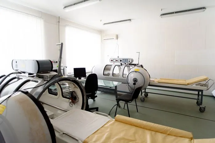
Hyperbaric Oxygen Therapy (HBOT): HBOT involves the administration of 100% oxygen at increased atmospheric pressure within a hyperbaric chamber. HBOT enhances tissue oxygenation, promotes angiogenesis, and reduces tissue hypoxia, improving wound healing outcomes in necrotic wounds.
Bioengineered Skin Substitutes: Bioengineered skin substitutes composed of living cells or synthetic biomaterials can promote wound healing in necrotic wounds. These substitutes provide a scaffold for cellular ingrowth, support tissue regeneration, and enhance wound closure in complex wounds.
Implementing a combination of these treatment modalities tailored to the necrotic wound's specific characteristics and underlying etiology is essential for achieving optimal outcomes and promoting tissue repair and regeneration. Close monitoring and regular reassessment of the wound are necessary to evaluate treatment efficacy, identify complications, and adjust the management plan accordingly.
Conclusion
Effective management of necrotic wounds requires a comprehensive understanding of the underlying mechanisms of necrosis and a tailored approach to treatment. Necrotic wounds pose significant challenges to wound healing due to non-viable tissue and impaired tissue perfusion. Treatment strategies for necrotic wounds include debridement techniques to remove necrotic tissue, appropriate wound dressings, topical treatments to promote healing, surgical interventions for severe cases, and advanced therapies such as negative pressure wound therapy and hyperbaric oxygen therapy. By implementing a multidisciplinary approach and combining these treatment modalities, healthcare providers can optimize wound healing outcomes and improve the quality of life for patients with necrotic wounds. Regular monitoring and reassessment of the wound are essential to evaluate treatment efficacy and ensure timely intervention when necessary.

.webp)
.avif)